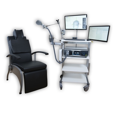
MotorMap TMS complete solution
This complete solution contains the diagnostic TMS + EMG setup from Neurosoft, the Neural Navigator Motor Mapping software and tracking unit, and the reclining patient chair. You do not need anything else to start mapping muscles with TMS and visualize evoked muscle EMG response on the brain surface. The TMS, EMG and navigation solution is a co-development of Neurosoft and Brain Science Tools BV, and integration is seamless. You do not need trigger cables or complicated configurations, this solution just works. This solution is cleared by the FDA for general purpose motor mapping in clinical use in the USA, CE certified for clinical use in the EU, and cleared for clinical use in Australia and Brazil
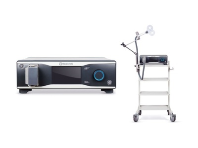
Neurosoft Neuro-MS monophasic TMS unit
High power monophasic pulse
Thanks to the 2800 V capacitor, the Neuro-MS TMS unit fires more powerful monophasic pulses. It ensures good motor response at low intensities. The peak magnetic field of Neuro-MS/D reaches 4 T, one of the highest values in the industry.
Pulse shape
The shape of the TMS pulse generated by this TMS stimulator is monophasic. Monophasic pulse shapes are better suited for diagnostic application of TMS, as the ensuing motor evoked potential signal is cleaner and easier to interpret.
Delayed charging mode
While working together with digital EMG system a delay for the capacitor charge start is set in the single pulse mode to avoid noise influence from the power supply unit during the acquisition. The range of the delay is 0–9.9 seconds.
Connection to computer via USB port
Neuro-MS/D can be used together with neurophysiological systems made by Neurosoft. Just connect the magnetic stimulator to the computer with installed EMG or EEG software via an USB port.
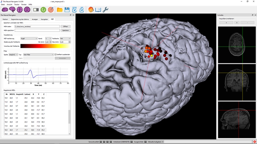
Neural Navigator MotorMap
Features & performance
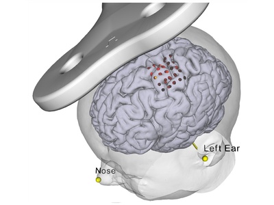
- Precision: The Neural Navigator can target brain areas indicated on an MRI scan with a precision of at least 4 mm (Neggers et al, 2004).
- Tracking technology: electromagnetic tracking using highly robust DC pulsed EM tracking (a technique often used in neurosurgical neuronavigation). Precision better than 1mm.
- MRI scans needed: straightforward T1 weighted MRI scan at around 1mm cubic resolution.
- Formats supported: DICOM, Nifti. Also supports fMRI activation overlays and targeting.
- Built in robust semi-automatic MRI scan segmentation.
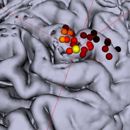
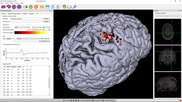
- Visualizes stimulation locations and evoked MEP amplitude or latency
- MEPs can be filtered, thresholded and manipulated in many other ways, and saved to disk to be loaded at a later time point for inspection or further analysis in martlab, python, etc.
- Actual 3D coil models for most Neurosoft coils built into the software.
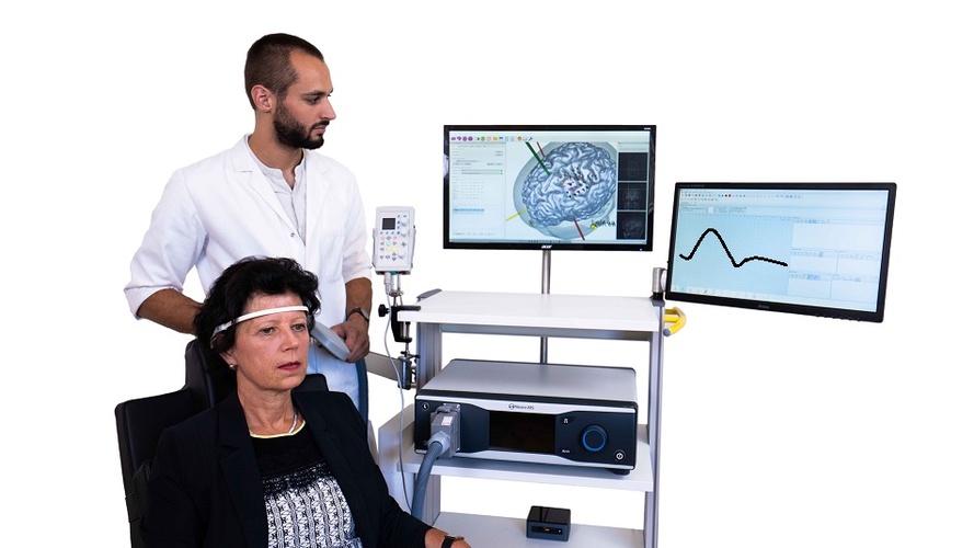
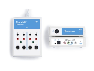
Neuro-MEP-Micro EMG system
- 2 channels are optimized to perform quickly motor and sensory conduction tests, needle EMG portable, can operate with notebook
- All-in-one: stimulators, acquisition channels, controls, display
- High acquisition quality: sampling rate – up to 100 kH electrical stimulator with ultra-fast switching between two outputs
Delivery set of complete system
- Neurosoft Neuro-MS monophasic TMS unit
- Figure-of-eight cooled coil 100 mm FEC-02-100-C
- Flexible arm for coil positioning
- Special trolley with 4 shelves, arm for EM transmitter and arm for 2nd screen, side mount for tracking system and coil arm.
- Coil holder , trolley or wall mounted
- The Neural Navigator software suite "MotorMap"
- The BrainTRAK neuronavigation position tracker with 4 tracking sensors and magnetic field transmitter.
- Plastic hand-held pointer for measuring facial landmarks and a remote control for navigation.
- A sturdy suitcase with compartments fitting all navigator components, allowing easy and safe transportation and storage
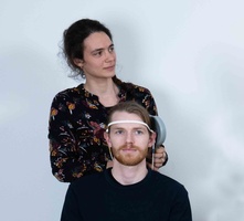
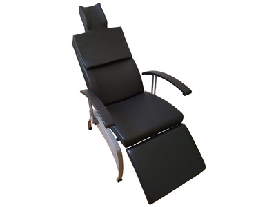
- A navigation compatible reclining comfortable TMS treatment chair.
- Technical and user manuals
- Neuro-MEP software to control TMS equipment from PC
- All required cabling
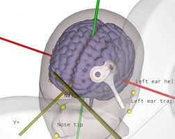
Reproducible coil placement using MNI
The neural navigator supports accurate reproduction of TMS coil positions using an MNI template. Similar to the standard navigation procedure, facial landmarks of the MNI template are captured on the participant's head. Next, the neural navigator adjusts the MNI template based on the geometry of the participant's head. MNI targets, …
TMS motor mapping in stroke
Transcranial magnetic stimulation (TMS) can be used to non-invasively induce a current and generate neuronal activity within the primary motor cortex. The neuronal activation travels through the corticospinal tract and the peripheral nervous system to finally elicit activity in various muscles, depending on the exact location of stimulation. The electrical …
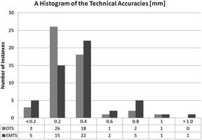
EM versus optical tracking in neuronavigation
During neuronavigation, the 3D position and angle of the TMS coil, a digitizing stylus and the head of the patient typically must be known at all times. Position tracking can be performed either using optical tracking with large cameras, or electromagnetic (EM) tracking based on a DC pulsed magnetic field …
Theta burst stimulation (TBS)
rTMS treatment protocols continue to be investigated and improved. New patterned rTMS protocols, where short high frequency trains of TMS pulses are are interleaved with really short breaks, are referred to as Theta Burst Stimulation (TBS), intermittent TBS (iTBS) and continues TBS (cTBS) with frequencies up to 100Hz for the …