TMS motor mapping in stroke
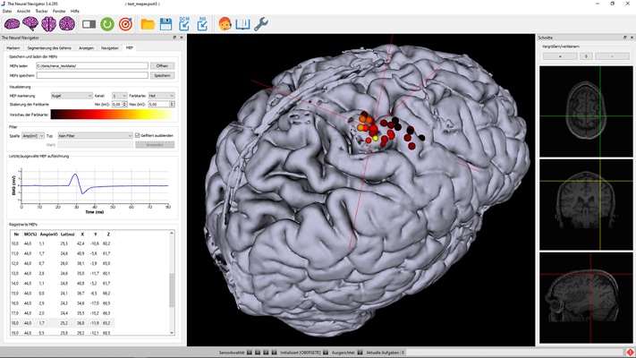
Transcranial magnetic stimulation (TMS) can be used to non-invasively induce a current and generate neuronal activity within the primary motor cortex. The neuronal activation travels through the corticospinal tract and the peripheral nervous system to finally elicit activity in various muscles, depending on the exact location of stimulation. The electrical activity in the muscle evoked by the TMS pulse on the motor cortex is called a motor-evoked potential (MEP) and can be measured using electromyography (EMG). TMS in combination with MRI-guided neuronavigation can then be used to identify parts of the cortex which are responsible for activity in specific muscles.
By stimulating a grid of targets in the motor-eloquent cortex, a map can be generated which represents the functional motor area of a certain muscle or muscle groups. From the functional motor area, several variables can be extracted, like the center of gravity and the surface area. These variables can be of value in the monitoring of recovery of motor function after stroke. For example, longitudinal changes in the spatial representation of the motor-eloquent cortex can be indicative of neuroplastic changes in the motor network which underly motor function recovery. Furthermore, the TMS motor maps provide information on the functional integrity of the motor cortex and the conduction time of the pathways.
We offer complete solutions with an MRI guided navigation system, monophasic TMS units and 4 channel EMG, optimized for MEP motor mapping, that fulfill all typical requirements.